
Your body contains about four grams of iron, roughly the weight of a small nail. Most of it sits quietly in your red blood cells, shuttling oxygen around your body. But iron has a darker side: when cellular controls fail, this essential metal becomes a weapon that cells use to destroy themselves in a process called ferroptosis.
What is iron chemistry in cells
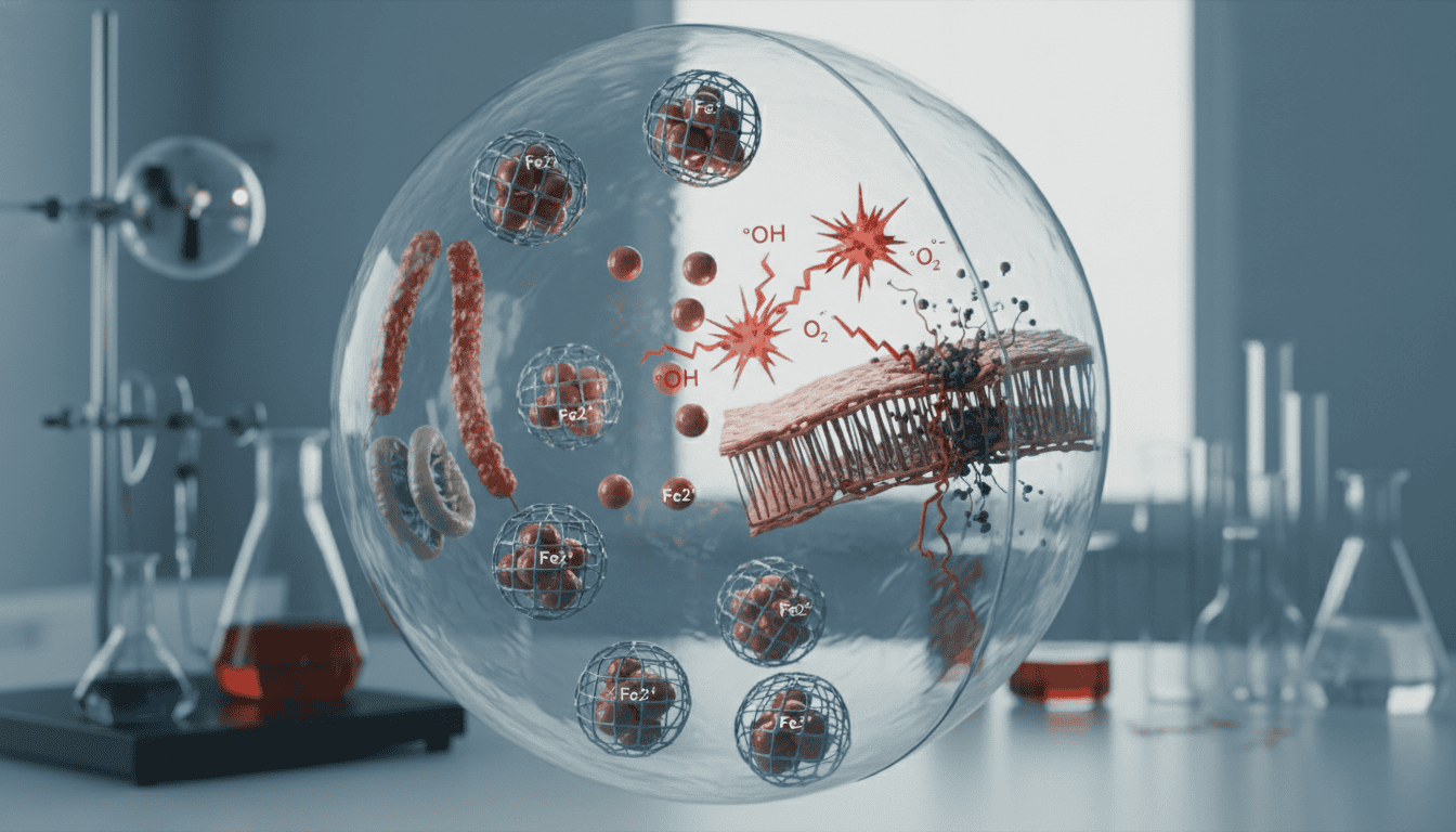
Iron exists in two main forms inside cells: ferrous (Fe2+) and ferric (Fe3+). Think of these as iron’s two personalities. Ferrous iron readily gives up electrons, making it perfect for biological reactions but also dangerously reactive. Ferric iron holds onto its electrons more tightly.
Cells store most of their iron inside a protein cage called ferritin. This keeps iron safe but accessible. When cells need iron for essential processes like DNA synthesis or energy production, they release it from storage. The catch? Free iron in the presence of hydrogen peroxide creates hydroxyl radicals through the Fenton reaction. These radicals attack cellular membranes like molecular scissors cutting through plastic wrap.
Ferroptosis represents a distinct form of programmed cell death driven entirely by iron-dependent lipid peroxidation. Unlike apoptosis, which cells execute through protein machinery, ferroptosis happens when iron chemistry overwhelms cellular defences. The cell’s membranes literally fall apart as iron-generated radicals chew through their fatty components.
What the research shows
Scientists first recognised ferroptosis in 2012 when they noticed certain cancer cells dying in an unusual way. The cells weren’t showing typical signs of apoptosis. Instead, their mitochondria were shrinking and their membranes were breaking down.
Research revealed that ferroptosis requires three key ingredients: iron, oxygen, and polyunsaturated fatty acids in cell membranes. Remove any one of these components and the process stops. Iron chelators that bind and sequester iron completely prevent ferroptosis. So do antioxidants that neutralise the radical chain reactions.
The process follows a predictable sequence. First, cellular antioxidant defences become overwhelmed or depleted. Then iron-catalysed reactions begin attacking membrane lipids, particularly those containing multiple double bonds. These damaged lipids create more radicals in a self-perpetuating cycle. Eventually, cellular membranes become so compromised that the cell dies.
Researchers have identified ferroptosis in neurons affected by stroke, kidney cells damaged by toxins, and various cancer cells under metabolic stress. The iron-dependent nature of the death pathway appears across many different cell types and disease contexts.
Why cells need this mechanism
Ferroptosis might seem purely destructive, but evolution rarely preserves mechanisms without purpose. This form of cell death likely serves as a cellular quality control system. When cells accumulate too much oxidative damage or lose their ability to maintain iron homeostasis, ferroptosis eliminates them before they can cause broader tissue damage.
The iron dependence makes biological sense. Iron-rich cells often have high metabolic activity, making them more prone to generating reactive oxygen species. If these cells lose their antioxidant defences, they become ticking time bombs. Ferroptosis provides a way to remove them quickly.
Cancer cells frequently show altered iron metabolism, accumulating more iron than healthy cells. This makes them potentially more vulnerable to ferroptosis, which researchers are exploring as a therapeutic target. The same iron dependence that makes cancer cells aggressive might also be their weakness.
What affects iron chemistry and ferroptosis
Diet influences cellular iron levels, but the body tightly regulates iron absorption. People with genetic iron overload conditions like haemochromatosis have cells that are more susceptible to ferroptosis. Conversely, iron deficiency can protect against ferroptosis but impairs many other cellular functions.
Antioxidant systems strongly influence ferroptosis susceptibility. The enzyme glutathione peroxidase 4 (GPX4) serves as a key defence, specifically neutralising lipid peroxides before they can propagate. Cells with robust GPX4 activity resist ferroptosis even under iron-rich conditions.
Inflammation affects both iron distribution and antioxidant defences. Chronic inflammatory conditions often involve dysregulated iron metabolism, with immune cells hoarding iron and tissues becoming iron-depleted. This redistribution can alter ferroptosis susceptibility across different tissues.
Age appears to influence ferroptosis sensitivity. Older cells often have accumulated more iron and show declining antioxidant capacity. This combination potentially makes them more vulnerable to iron-driven cell death, though researchers are still mapping these relationships.
What remains unknown
Scientists still don’t fully understand how cells decide between different forms of programmed death. What tips the balance toward ferroptosis rather than apoptosis? The signalling pathways that initiate ferroptosis remain partially mysterious.
The role of ferroptosis in healthy ageing versus disease progression needs clarification. Does ferroptosis contribute to age-related tissue decline, or does it protect against worse outcomes by eliminating damaged cells? The answer likely varies by tissue and context.
Researchers are working to identify reliable biomarkers for ferroptosis in living systems. Most current detection methods require laboratory analysis of cells or tissues. Finding ways to measure ferroptosis activity in real-time would advance both research and potential therapeutic applications.
The relationship between ferroptosis and other cellular stress responses remains unclear. How do cells integrate signals about iron levels, oxidative stress, and metabolic status to determine their fate? This decision-making process involves complex molecular networks that scientists are still deciphering.
Iron chemistry reveals how the same elements that enable life can also orchestrate death. This duality reflects a broader principle in biology: our most essential systems often operate closest to the edge of chaos. Understanding ferroptosis illuminates how cells navigate the narrow space between using iron’s reactive power and being destroyed by it. As research continues, this iron-dependent death pathway may reveal new insights into how cellular quality control systems maintain the delicate balance that keeps us alive.
Matt Elliott is the editor of Redox News Today, an independent publication covering peer-reviewed research on cellular health, redox signalling, and related biomedical science.


