Every time someone takes a drag on a cigarette, their lungs absorb more than 4,000 chemical compounds and 10^15 free radicals. That’s a quadrillion reactive molecules hitting lung tissue in seconds. But the damage doesn’t stop there. These oxidative assaults trigger a cascade that reaches all the way to the brain, where iron begins accumulating in neurons through pathways scientists are only now beginning to understand.
What is lung-brain oxidative stress signalling
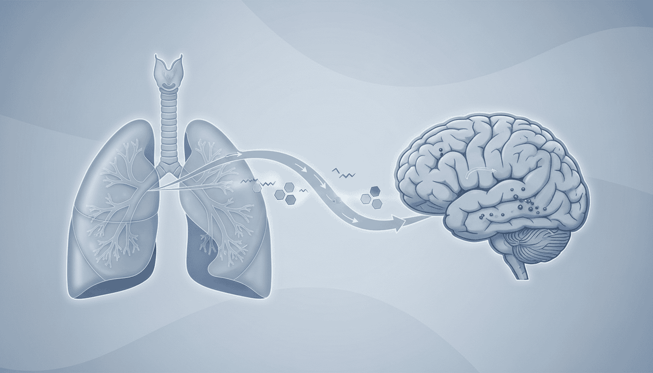
Your lungs and brain communicate through multiple biochemical pathways, and cigarette smoke disrupts several of them simultaneously. When tobacco smoke hits lung tissue, it immediately generates massive amounts of reactive oxygen species. These free radicals overwhelm local antioxidant defences within minutes.
The lung responds by releasing inflammatory signals. Cytokines like TNF-alpha and interleukin-6 enter the bloodstream and travel to the brain. Once there, they activate microglia, the brain’s immune cells, which start producing their own reactive oxygen species. This creates a second wave of oxidative stress, but now inside neural tissue.
Meanwhile, the blood-brain barrier becomes more permeable under oxidative assault. Normally this barrier keeps potentially harmful substances in blood from entering brain tissue. Cigarette smoke metabolites can now cross more easily, bringing oxidative damage directly to neurons.
What the research shows
Studies using brain imaging have revealed something unexpected: people who smoke show increased iron deposits in specific brain regions, particularly the substantia nigra and putamen. These areas are rich in dopamine-producing neurons.
Animal studies provide the mechanistic details. When researchers expose mice to cigarette smoke, they see disrupted iron regulation within weeks. The protein ferritin, which normally stores iron safely inside cells, becomes overwhelmed. Free iron accumulates in neurons, where it catalyses the production of hydroxyl radicals through the Fenton reaction.
Brain tissue from smokers shows elevated levels of lipid peroxidation markers. These are the molecular fingerprints left behind when free radicals attack cell membranes. The pattern suggests ongoing oxidative damage that persists even during periods when people aren’t actively smoking.
Research has also tracked how quickly this lung-brain connection develops. Inflammatory markers appear in cerebrospinal fluid within hours of smoke exposure. Iron accumulation takes longer, building up over months and years of regular smoking.
Why cells need iron regulation
Iron sits at the centre of cellular energy production. Every mitochondrion needs iron-containing enzymes to generate ATP through the electron transport chain. Your brain, which uses about 20% of your body’s energy despite being only 2% of your weight, requires especially tight iron control.
But iron’s chemical properties that make it useful also make it dangerous. The same ability to transfer electrons that powers cellular respiration also generates devastating free radicals when iron isn’t properly contained. Evolution solved this problem with sophisticated storage and transport systems.
Neurons have particularly vulnerable fatty membranes loaded with polyunsaturated fats. When free iron meets these membranes, it triggers lipid peroxidation cascades that can damage large sections of cell membrane. A single iron atom can potentially destroy hundreds of lipid molecules through chain reactions.
The brain also lacks some of the antioxidant enzymes found in other organs. This makes proper iron storage even more critical for neural survival.
What affects lung-brain oxidative stress
Cigarette intensity matters more than most people realise. Light smokers show measurable brain iron accumulation, but heavy smokers develop it faster and in more brain regions. The relationship isn’t linear though. Even occasional smoking appears to trigger some degree of oxidative signalling between lungs and brain.
Age amplifies the effects significantly. Older smokers show more dramatic iron accumulation than younger ones exposed to similar amounts of smoke. This likely reflects declining antioxidant capacity and slower cellular repair processes that come with ageing.
Genetic variations in iron metabolism genes influence individual responses. People with certain variants of the HFE gene, associated with iron overload conditions, appear more susceptible to smoking-related brain iron accumulation.
Air pollution creates additive effects. People who smoke and live in areas with high particulate matter show enhanced oxidative stress markers compared to smokers in cleaner environments. Both cigarette smoke and air pollution generate similar types of free radicals.
Diet plays a role too. Antioxidant nutrients like vitamin C and E can partially buffer the oxidative assault, though they can’t completely prevent it. Iron intake through food or supplements may worsen the accumulation in smokers.
What remains unknown
Scientists still don’t fully understand why some brain regions accumulate iron more readily than others in smokers. The substantia nigra and putamen are consistently affected, but researchers haven’t identified what makes these areas particularly vulnerable.
The reversibility question remains partly unanswered. Some studies suggest brain iron levels can decrease after smoking cessation, but the timeline and completeness of this recovery varies dramatically between individuals. Whether the oxidative damage to neurons can be fully repaired is still unclear.
Researchers are also investigating whether other forms of inhaled oxidative stress, like vaping or marijuana smoke, trigger similar lung-brain pathways. Early studies suggest they might, but the specific patterns of iron accumulation appear different.
The interaction between smoking-induced iron accumulation and neurodegenerative diseases represents another frontier. While associations exist, the exact causal relationships remain under investigation.
This research reveals how oxidative stress creates far-reaching connections between organ systems. The lung-brain axis represents just one example of how localised cellular damage can trigger systemic responses. Understanding these pathways helps explain why oxidative stress, whether from smoking or other sources, rarely affects just one part of the body. Instead, it creates cascading effects that can influence cellular function throughout multiple organs, often in ways that take years to fully manifest.

Matt Elliott is the editor of Redox News Today, an independent publication covering peer-reviewed research on cellular health, redox signalling, and related biomedical science.