
Your muscle cells can pack in thousands of mitochondria when you’re training hard, but barely a few hundred when you’re sedentary. The difference isn’t magic. It’s a single protein called PGC-1α that acts like a construction foreman, deciding when your cells need more power plants and then building them from scratch.
What is PGC-1α
PGC-1α stands for peroxisome proliferator-activated receptor gamma coactivator 1-alpha, though thankfully scientists just call it PGC-1α. Think of it as your cell’s energy consultant.
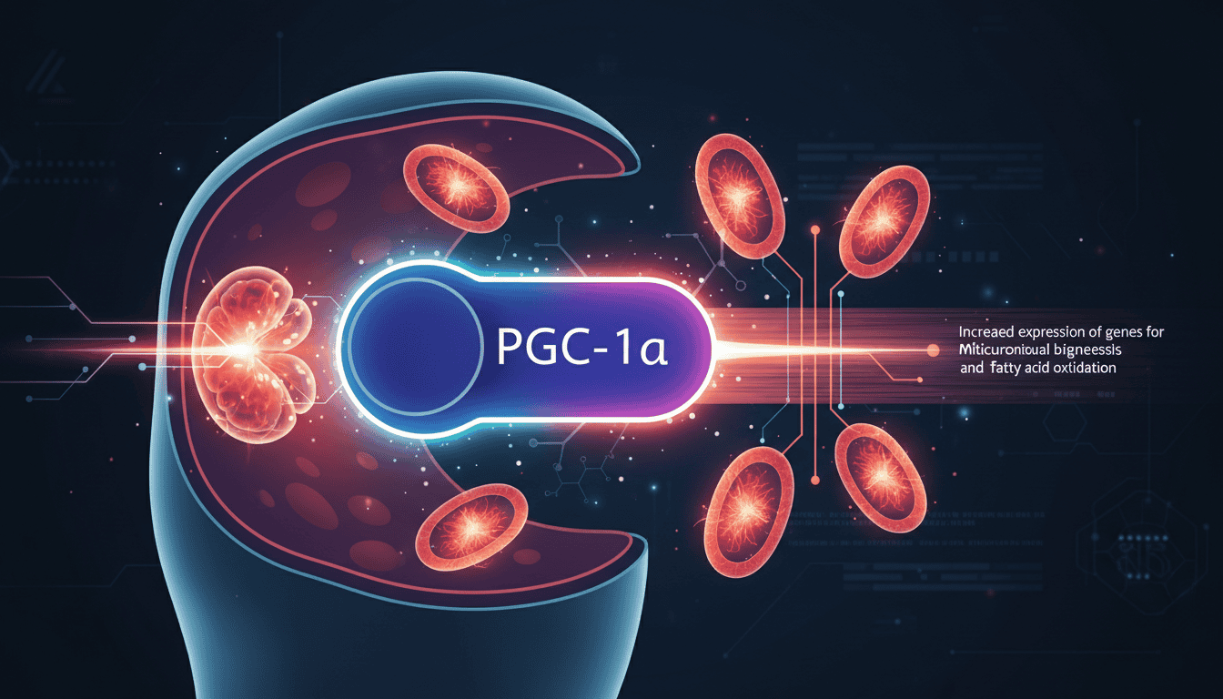
When your cells sense they need more energy production capacity, PGC-1α springs into action. It doesn’t make mitochondria directly. Instead, it coordinates with dozens of other proteins to switch on the genes needed for mitochondrial biogenesis. This process involves creating new mitochondrial proteins, importing them into existing mitochondria, and eventually splitting those mitochondria into two functional units.
PGC-1α also fine-tunes how efficiently your existing mitochondria work. It boosts the production of enzymes in the electron transport chain, the molecular assembly line that converts nutrients into ATP. Without adequate PGC-1α activity, your mitochondria become sluggish and fewer in number.
The protein doesn’t work alone. It partners with transcription factors like NRF1 and NRF2, which bind to DNA and activate specific genes. PGC-1α essentially tells these factors which genes to turn on and how strongly to activate them.
What the research shows
Studies using muscle biopsies reveal that endurance athletes have dramatically higher levels of PGC-1α compared to sedentary individuals. Their muscle cells also contain up to twice as many mitochondria, and each mitochondrion produces ATP more efficiently.
Laboratory experiments show that artificially increasing PGC-1α levels in muscle cells triggers a cascade of mitochondrial growth. Within days, researchers observe new mitochondrial proteins appearing and existing mitochondria beginning to divide. The cells start consuming oxygen more rapidly and producing more ATP.
Research in mice demonstrates that animals engineered to overproduce PGC-1α develop what scientists call a “super-endurance” phenotype. Their muscles contain dense networks of mitochondria and resist fatigue during prolonged exercise. Conversely, mice lacking PGC-1α show reduced mitochondrial numbers and poor exercise capacity.
Studies on ageing reveal a consistent pattern: PGC-1α levels decline with age across multiple tissues. This decline correlates with reduced mitochondrial function and the metabolic changes associated with getting older. Tissue samples from elderly individuals show fewer mitochondria per cell and reduced expression of genes involved in energy metabolism.
Cold exposure research provides another angle. When mammals are exposed to cold temperatures, PGC-1α levels spike in brown fat tissue. This drives the production of mitochondria specialised for heat generation rather than ATP production, helping maintain body temperature.
Why cells need this
Energy demands fluctuate wildly throughout life. A marathon runner needs vastly more cellular energy production than someone sitting at a desk. But building mitochondria requires significant resources and time.
PGC-1α solves this problem by providing a responsive system. When energy demands increase, cells can ramp up mitochondrial biogenesis. When demands decrease, they can scale back production and even remove excess mitochondria through autophagy.
This flexibility offers survival advantages. Animals that could quickly adapt their energy production capacity would outcompete those with fixed mitochondrial numbers. During food scarcity, reducing mitochondrial mass saves precious amino acids and other building materials. During abundance or high activity periods, increasing mitochondrial capacity enables better performance.
PGC-1α also coordinates mitochondrial function with other cellular processes. It influences genes involved in fat oxidation, glucose metabolism, and antioxidant defence. This integration ensures that energy production systems work in harmony rather than competing for resources.
What affects PGC-1α
Exercise stands out as the most potent stimulator of PGC-1α. Both endurance exercise and high-intensity interval training trigger increased PGC-1α expression, though through slightly different molecular pathways. The effect begins within hours of exercise and can persist for days.
Cold exposure activates PGC-1α, particularly in brown fat tissue but also in muscle. This response helps generate heat through increased mitochondrial activity. Regular cold exposure appears to maintain higher baseline PGC-1α levels.
Fasting and caloric restriction influence PGC-1α in complex ways. Short-term fasting often increases PGC-1α as cells adapt to limited fuel availability. However, prolonged severe restriction can eventually suppress it as the body conserves resources.
Sleep patterns affect PGC-1α regulation. Research shows that circadian rhythm disruption can interfere with normal PGC-1α cycling, potentially explaining why shift work and poor sleep correlate with metabolic problems.
Ageing consistently reduces PGC-1α levels across tissues. This decline appears to be both a cause and consequence of mitochondrial dysfunction in older adults. Various age-related factors, including chronic inflammation and hormonal changes, may contribute to this reduction.
What remains unknown
Scientists still debate exactly how cells sense when they need more mitochondria. Multiple signalling pathways can activate PGC-1α, but the relative importance of each pathway under different conditions remains unclear.
The relationship between PGC-1α and mitochondrial quality control presents another puzzle. While PGC-1α clearly drives mitochondrial biogenesis, researchers are still working out how it coordinates with systems that remove damaged mitochondria.
Individual variation in PGC-1α responses puzzles researchers. Some people show dramatic increases in PGC-1α after exercise, while others show modest changes. Genetic factors likely play a role, but environmental and epigenetic influences remain poorly understood.
The optimal levels of PGC-1α for different tissues and life stages aren’t established. While more PGC-1α generally correlates with better mitochondrial function, it’s unclear whether there’s an upper limit or optimal range for different physiological conditions.
Tissue-specific differences in PGC-1α regulation need more research. The protein behaves differently in muscle, liver, brain, and fat tissue, but the mechanisms behind these differences aren’t fully mapped.
PGC-1α represents one of biology’s elegant solutions to a fundamental challenge: how to match energy supply with demand in a changing world. As researchers continue mapping the intricate networks that control cellular energy production, PGC-1α stands as a reminder that even our most basic cellular functions rely on sophisticated regulatory systems that have been millions of years in the making.
Matt Elliott is the editor of Redox News Today, an independent publication covering peer-reviewed research on cellular health, redox signalling, and related biomedical science.


