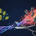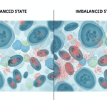
A fetus with severe anaemia faces a metabolic crisis. Oxygen delivery drops. Red blood cells dwindle. And inside struggling cells, a particular type of damage accelerates: oxidative stress, where unstable molecules called free radicals overwhelm the cell’s repair systems. What happens next is a story of cellular survival that hinges on a single protein, one so important that scientists call it “the guardian of the genome.”
What is oxidative stress and the p53 response
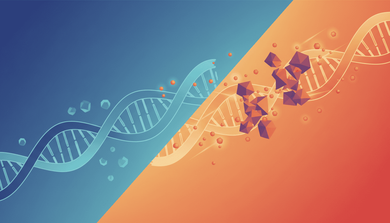
Oxidative stress isn’t mystical. It’s a chemical imbalance. Cells produce energy through metabolism, and that process generates free radicals as a byproduct. Normally, enzymes like catalase and superoxide dismutase neutralise these rogue molecules before they cause trouble. But when oxygen availability drops, as it does in severe fetal anaemia, cells struggle to keep up with radical production. The radicals start damaging DNA, proteins, and lipids.
This is where p53 enters. When cells detect this damage, p53 activates like a fire alarm. The protein accumulates in the cell nucleus, binds to specific DNA sequences, and switches on dozens of protective genes simultaneously. Some encode enzymes that repair oxidative damage. Others trigger cell cycle checkpoints that pause growth while repair happens. If damage is too severe, p53 can even initiate apoptosis, a programmed cell death that removes the damaged cell before it becomes dangerous.
In fetal anaemia, this response becomes matter of life and death. Erythroid progenitor cells, which are supposed to multiply rapidly to replace lost red blood cells, face a dilemma. Oxidative stress signals danger. p53 responds by putting the brakes on. The evolutionary logic is sound: better to slow production than to manufacture defective cells.
What the research shows
Studies examining fetal anaemia models have revealed how severely oxidative stress can compromise erythropoiesis, the process of red blood cell production. When researchers induce anaemia in fetal tissues or cultured erythroid cells, oxidative markers increase markedly. Hydrogen peroxide accumulates. Lipid peroxidation signatures appear. Protein oxidation rises.
Alongside these changes, p53 activation follows predictably. Nuclear p53 levels climb. Expression of p53 target genes surges, particularly those encoding antioxidant enzymes like peroxiredoxins and glutathione S-transferases. The cell is mounting a defence. But here’s the catch: p53 also upregulates genes that halt the cell cycle. In cells that should be dividing furiously to replace lost red blood cells, this creates a production bottleneck.
Research using p53-deficient models reveals the cost of losing this guardian. Cells without functional p53 do continue dividing under oxidative stress, but they accumulate mutations and chromosomal abnormalities. Some develop into aberrant cells. The temporary slowdown imposed by p53, it turns out, prevents a worse disaster downstream.
Tissue samples from cases of severe fetal anaemia show elevated p53 signalling in erythroid precursors. The activation pattern suggests cells are caught in a holding pattern: enough stress to trigger p53, but not enough resolution to allow division to resume. This can perpetuate anaemia even when the underlying cause persists.
Why cells need this defence mechanism
Evolution didn’t design p53 as a luxury. It’s a survival essential. Oxidative damage to DNA creates mutations. Mutations in actively dividing cells like blood precursors can lead to leukaemia or other blood cancers. A cell temporarily unable to divide is inconvenient. A cell dividing with a damaged genome is catastrophic.
In the context of fetal development, this trade-off makes particular sense. Fetal tissues are undergoing rapid expansion and differentiation. Speed matters. But accuracy matters more. A single malignant mutation in a blood-forming stem cell could establish a clone that expands throughout the fetus, causing haematologic malignancy later in life.
The p53 system essentially asks a cellular question: do we have the resources to divide safely? When oxidative stress signals resource depletion, p53 says no. It buys time for repair mechanisms to function. It’s a conservative strategy, one that sacrifices short-term production for long-term genomic integrity.
What affects oxidative stress and p53 activation
Several factors influence how severely oxidative stress develops in fetal anaemia. Maternal oxygen tension obviously matters. In pregnancies at high altitude or complicated by placental insufficiency, fetal oxygen availability drops further, intensifying oxidative stress in tissues already struggling with anaemia.
Nutritional status plays a role too. Antioxidant micronutrients like vitamin E, selenium, and zinc support the enzymes that neutralise free radicals. Deficiency in these nutrients weakens the cell’s first line of defence against oxidative damage, pushing the burden onto p53-mediated responses.
The severity and duration of anaemia itself matters. Chronic mild anaemia allows some adaptation. Acute severe anaemia creates a metabolic crisis where oxidative stress overwhelms repair capacity quickly. In fetal contexts, the developmental stage at which anaemia begins influences outcomes too. Earlier gestational exposure may trigger different compensatory responses than late-gestation complications.
Genetic variation in genes encoding antioxidant enzymes and p53 itself also influences the response. Some individuals have naturally higher basal levels of these protective systems. Others start with less buffer against oxidative damage.
What remains unknown
One unresolved question concerns the optimal duration of p53-mediated cell cycle arrest. How long should erythroid progenitors pause before resuming division? Too short and cells divide with unrepaired damage. Too long and anaemia worsens from insufficient red blood cell production. Research hasn’t established whether p53 signalling in this context is calibrated optimally or whether it sometimes errs too far toward caution.
Another gap concerns interactions between p53 and erythroid-specific transcription factors. The proteins that normally drive red blood cell maturation, like GATA1 and KLF1, likely interact with p53 signalling in ways researchers are still mapping. Understanding these crosstalk mechanisms might reveal why some fetal anaemias resolve despite ongoing oxidative stress, while others persist.
Researchers also lack detailed understanding of tissue-specific differences. Oxidative stress affects multiple fetal organs during anaemia, but p53 responses may vary substantially between erythroid cells, cardiomyocytes, and neuronal cells. Why some tissues tolerate oxidative stress better than others remains partly mysterious.
Finally, the long-term consequences of fetal oxidative stress exposure are incompletely characterised. Does experiencing oxidative stress during critical developmental windows programme cells differently? Do surviving red blood cells show functional differences? These questions matter for understanding whether fetal anaemia leaves lasting marks on cellular physiology.
The oxidative stress response in fetal anaemia illustrates a broader principle in cellular biology: stress detection and adaptation mechanisms exist in constant tension. Cells need to react quickly to danger, yet respond proportionally to avoid overreaction. The p53 system represents one elegant solution to that challenge, a protein that translates chemical distress signals into coordinated protective action. Understanding these defence pathways reveals not just how cells survive crisis, but how billions of years of evolution have wired survival into our molecular machinery.

Matt Elliott is the editor of Redox News Today, an independent publication covering peer-reviewed research on cellular health, redox signalling, and related biomedical science.