When a blood clot blocks an artery feeding the brain, neurons die within minutes. But the damage doesn’t stop there. The cells surrounding those dead neurons spring into action, and one of their responses is counterintuitive: they start manufacturing collagen, the same structural protein found in skin and tendons. This process, triggered by oxidative stress, shapes whether the brain can recover or suffers lasting harm.
What is astrocytic collagen production
Astrocytes are star-shaped support cells that outnumber neurons in the brain. They don’t fire action potentials, but they do almost everything else: they buffer chemicals, provide nutrients, clear waste, and regulate the brain’s immune response. During a stroke, when oxygen-starved tissue generates reactive oxygen species (ROS) in a desperate, chaotic burst, astrocytes detect this oxidative stress and shift into a different mode.
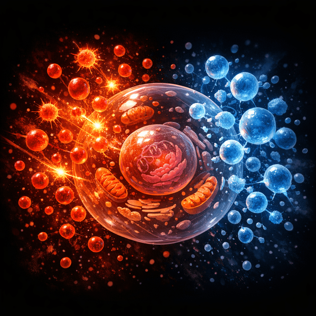
Oxidative stress occurs when the body produces more unstable molecules than it can neutralise. These ROS molecules, like superoxide and hydrogen peroxide, damage proteins, fats, and DNA. In the stroke brain, ROS levels spike dramatically. Astrocytes recognise this chemical signal and activate specific pathways that drive them to produce and secrete collagen, particularly type I and type IV collagen. This response involves signalling cascades that alter which genes the astrocytes express, essentially reprogramming them from housekeeping mode into builders.
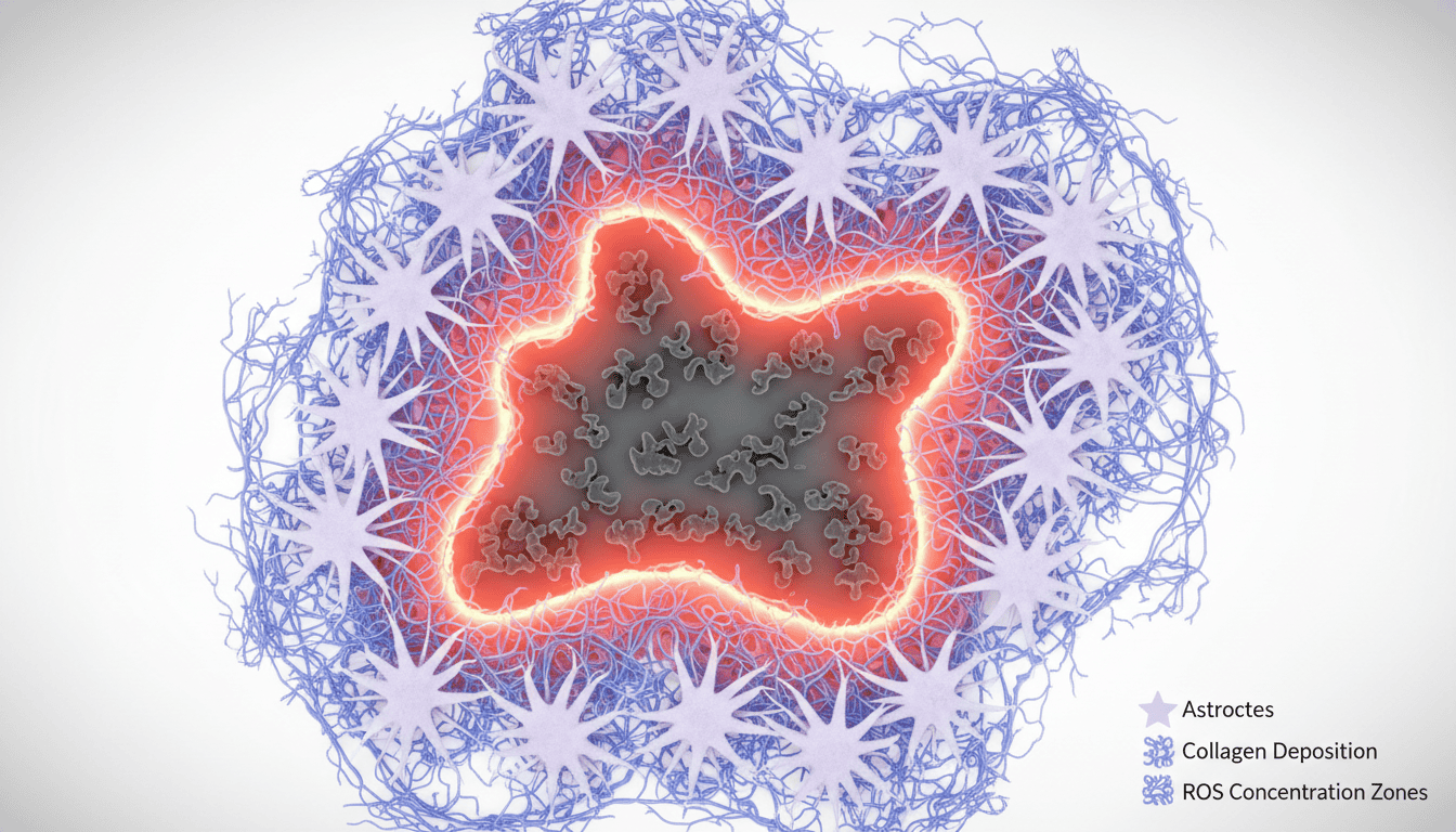
The collagen they produce becomes part of the scar tissue that forms around the damaged area. Think of it as the brain’s attempt to wall off the injury, creating a physical barrier that stabilises the tissue architecture.
What the research shows
Studies using animal models of ischaemic stroke have shown that oxidative stress directly correlates with increased collagen deposition in the perilesional region, the area surrounding the dead tissue. Researchers observed that when they blocked antioxidant defences experimentally, collagen production increased. Conversely, when they enhanced antioxidant activity, collagen accumulation decreased.
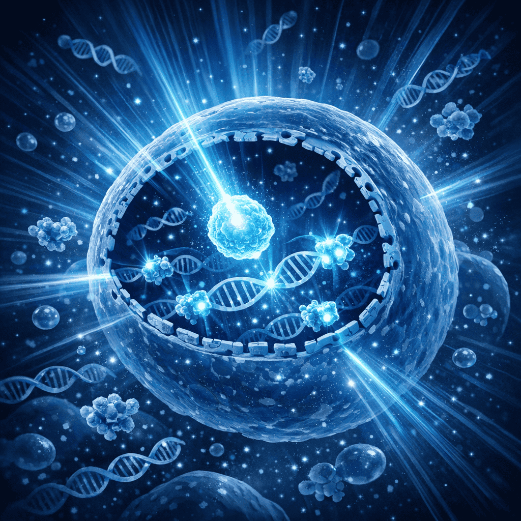
The mechanism involves several signalling pathways working in parallel. ROS activates transcription factors like NRF2 and NF-kappa B, which then switch on genes encoding collagen synthesis enzymes. Astrocytes also respond to inflammatory cytokines released by dying cells and activated immune cells, and these signals amplify the collagen-building response. Imaging studies show that this collagen remodelling starts within hours of the stroke and continues for days and weeks.
One striking finding is that the extent of collagen deposition varies depending on how severe the oxidative stress becomes. Moderate oxidative stress triggers a measured collagen response. Extreme stress can lead to excessive collagen accumulation, which paradoxically harms recovery because dense scar tissue restricts the rewiring of surviving neural circuits. This suggests the relationship between ROS and collagen production isn’t simply linear.
Why cells need this
From an evolutionary perspective, astrocytic collagen production makes sense as damage containment. When tissue is injured, the structural matrix falls apart. Collagen provides scaffolding that prevents further tissue collapse and loss of blood vessel integrity. In the hours after a stroke, this might prevent the injury from expanding.
Collagen also provides physical support for axons attempting to regrow and reconnect. The scar tissue, though often portrayed as purely obstructive, can also serve as a substrate for cell migration and neuronal sprouting. The astrocytes themselves embed within this collagen, and they use it to structure their own networks of connexions with other cells.
The ROS signalling component adds another layer. Oxidative stress isn’t just damage; it’s information. Cells interpret high ROS levels as an emergency signal meaning “tissue is dying here, mobilise structural reinforcement.” The astrocytes respond with collagen production as part of a broader damage-response programme that also includes inflammatory signalling and tissue remodelling.
What affects astrocytic collagen production
Age significantly influences this process. Older brains show exaggerated collagen responses to the same degree of stroke injury, partly because their antioxidant defences decline with time. Aged astrocytes produce collagen less efficiently but generate more reactive oxygen species, creating a mismatch between the threat and the response capacity.
Metabolic state matters too. Brains chronically exposed to glucose dysregulation or high oxidative stress before a stroke respond differently than metabolically healthy brains. Chronic oxidative stress can exhaust astrocytic antioxidant reserves, changing how they respond acutely.
Sex differences emerge in the research as well. Male and female astrocytes show different sensitivity to ROS signalling and different rates of collagen synthesis following oxidative stress. This likely reflects differences in their baseline antioxidant enzyme expression and how oestrogen modulates oxidative stress responses.
Environmental factors like prior exercise, diet quality, and sleep history influence the baseline antioxidant capacity of brain cells and therefore shape their response to an acute stroke. Tissue hypoxia severity determines ROS magnitude, which in turn sets the intensity of the collagen production signal.
What remains unknown
The field still doesn’t fully understand why some collagen deposition aids recovery while excessive accumulation hinders it. The threshold between beneficial and harmful scar formation remains unclear, and we lack reliable markers to predict whether a given patient’s astrocytic response will be protective or problematic.
Researchers are uncertain about the timeline. How long should collagen accumulation continue before it becomes counterproductive? Can the timing of collagen formation be therapeutically optimised? Does the composition of the collagen matter, or only its quantity?
The interaction between astrocytic collagen production and other cells also requires more work. How do microglia and oligodendrocyte precursors respond to this collagen scaffold? Do they facilitate or frustrate recovery?
Finally, human data remains sparse. Most research uses rodent models with relatively uniform genetics and controlled environments. Whether these findings translate to the heterogeneous human brain, with its individual variation in genetics, prior health status, and stroke characteristics, remains an open question.
Understanding how oxidative stress drives astrocytic collagen production during stroke illustrates a fundamental principle in cellular biology: the boundary between damage and repair is often blurry. Cells don’t simply turn on a damage-response switch; they integrate multiple signals and produce graded responses calibrated to threat severity. The astrocyte’s collagen production is one example of how cells attempt to stabilise their environment when chaos erupts. Mapping these responses in detail might eventually reveal points where the outcome could be shifted in a more favourable direction, though that possibility remains speculative until far more is understood about the system’s operating principles.

Matt Elliott is the editor of Redox News Today, an independent publication covering peer-reviewed research on cellular health, redox signalling, and related biomedical science.