Your bones are constantly being dismantled and rebuilt. Every day, specialised cells called osteoclasts tear down old bone tissue while osteoblasts lay down new material to replace it. This process sounds exhausting because it is. Building bone requires enormous amounts of energy, and that energy comes almost entirely from mitochondria, the cellular power plants. When mitochondrial function falters, bone formation stumbles too. Understanding this connection is reshaping how researchers think about bone health and repair.
What is mitochondrial bone metabolism
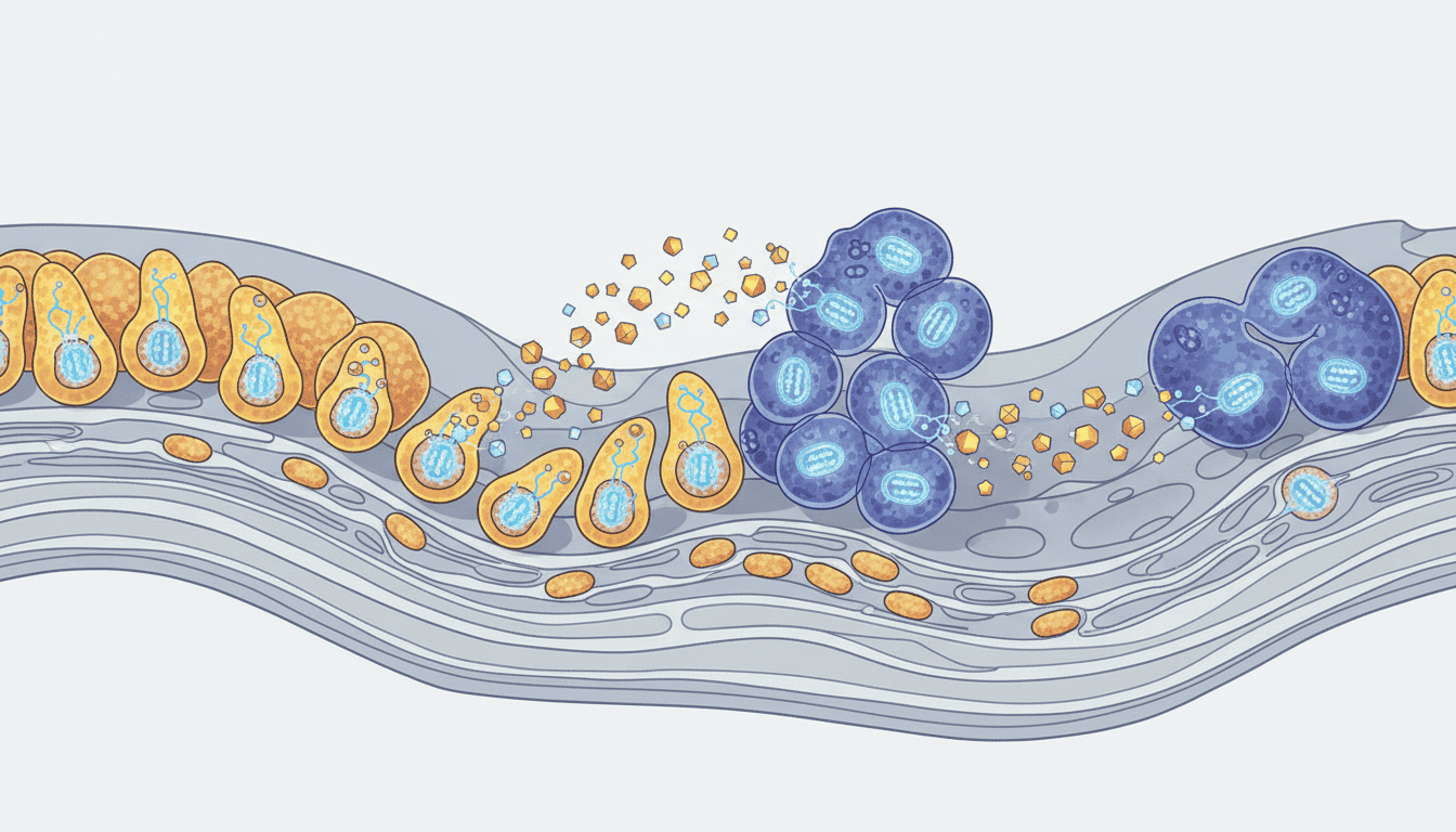
Bone is not static. It is a living tissue constantly remodelling itself, a process that demands metabolic fuel. Osteoblasts, the cells responsible for laying down new bone matrix, are metabolically ravenous. They need ATP, the cellular energy currency, to fuel the synthesis of collagen and mineralisation of the bone matrix. But they also need something else: reactive oxygen species (ROS) in carefully controlled amounts.
Mitochondria produce both ATP and ROS as byproducts of respiration. The relationship is delicate. Too little energy and osteoblasts cannot build bone efficiently. Too much oxidative stress and the cells become damaged. Osteoclasts, the cells that break down bone, have their own mitochondrial demands. When mitochondria in these cells work properly, bone remodelling stays balanced. When they do not, the whole system shifts out of equilibrium.
This is not just about energy availability, though that matters enormously. Mitochondria also regulate calcium handling, control cell death pathways, and influence the signalling molecules that tell bone cells when to activate or rest. The organelle sits at the centre of bone metabolism in ways researchers are still mapping out.
What the research shows
Studies examining osteoblasts in culture reveal something striking: when mitochondrial function is experimentally impaired, bone-forming capacity drops dramatically. Researchers observe reduced alkaline phosphatase activity, decreased collagen synthesis, and lower mineralisation rates. The cells are trying to build bone but lack the energetic resources to do it properly.
Animal models tell a similar story. Mice engineered to have defective mitochondrial function in osteoblasts develop weaker bones with reduced density and structural integrity. Their fracture healing is delayed and incomplete. The bones do regenerate eventually, but more slowly and less effectively than in animals with normal mitochondrial activity.
Osteoclasts show an inverse pattern. These bone-resorbing cells are also heavily dependent on mitochondrial ATP production. When their mitochondria are compromised, they become less efficient at breaking down bone, which might sound beneficial until you realise it disrupts the finely tuned remodelling cycle. The balance between building and breaking down is what keeps bone healthy and adaptive.
Research on ageing bone tissue reveals something important: mitochondrial function declines with age in osteoblasts. These cells accumulate damaged mitochondria, produce less ATP, and show increased oxidative stress. This correlates directly with age-related declines in bone formation and increased fracture risk. Young bone cells have more abundant, healthier mitochondria. Older ones do not.
Why cells need this
The energetic demands of bone formation are enormous by cellular standards. Manufacturing and secreting the bone matrix proteins, particularly collagen, requires sustained ATP production. Mineralisation, the process where hydroxyapatite crystals integrate into the matrix, demands continuous energy input. A single osteoblast might produce hundreds of micrograms of bone matrix material during its lifetime. That cannot happen without robust mitochondrial support.
Beyond raw energy, mitochondria regulate intracellular calcium signalling, which directly controls when osteoblasts activate their bone-building programmes. They also manage the oxidative environment within cells. Bone formation itself generates ROS as a signalling molecule, not just as waste. Mitochondria help keep this signalling in the productive range, neither too high nor too low.
Evolution preserved this tight coupling between mitochondrial function and bone metabolism because bones must be responsive to mechanical stress and systemic demands. A skeleton with reliable, adaptive remodelling is far more valuable than one locked into a fixed architecture. That responsiveness requires metabolic flexibility and energy security, both provided by healthy mitochondria.
What affects mitochondrial bone function
Physical activity directly influences mitochondrial function in bone cells. Exercise increases ATP demand throughout the body, which stimulates mitochondrial biogenesis in multiple tissues including bone. More and healthier mitochondria follow. This is one mechanism through which mechanical loading strengthens bone at the cellular level.
Nutrition shapes mitochondrial capacity. Adequate carbohydrates and fats provide substrate for ATP production. Micronutrients like magnesium, phosphate, and B vitamins support mitochondrial enzyme function. Antioxidants help manage the oxidative environment. A diet lacking in these elements compromises mitochondrial performance in osteoblasts.
Ageing itself remains the largest modifier. Mitochondrial DNA accumulates mutations over time. The cellular quality control systems that remove damaged mitochondria become less efficient. Osteoblasts in older individuals have fewer functional mitochondria per cell and higher baseline oxidative stress. This partly explains why bone formation slows with age and why fracture repair becomes less reliable.
Hormonal status affects mitochondrial biogenesis in bone cells. Oestrogen signalling influences mitochondrial gene expression in osteoblasts. This helps explain why postmenopausal declines in oestrogen correlate with reduced bone formation capacity. Other hormones like thyroid hormone and parathyroid hormone also modulate mitochondrial activity in bone cells.
What remains unknown
Researchers still cannot fully explain how mitochondrial ROS production contributes to bone formation. The relationship is definitely not simple. Some ROS is required for osteoblast differentiation and function. Too much causes oxidative damage. Where exactly the optimal level sits, and how that optimum shifts with age or disease, remains unclear.
The communication pathways between mitochondria and the genes controlling bone formation are not completely mapped. How do mitochondria signal their energetic state to the cell nucleus? What triggers increased mitochondrial biogenesis specifically in osteoblasts during bone repair? These questions matter for understanding bone regeneration.
Whether interventions that directly improve mitochondrial function could enhance bone regeneration in humans remains untested at scale. Animal studies suggest promise, but translating laboratory findings to clinical settings is never straightforward. The bone cells in a living person face constraints and pressures that isolated cells do not.
The relationship between mitochondrial function and osteoclast activity during fracture healing also needs clarification. We know osteoclasts require functional mitochondria, but how their mitochondrial activity coordinates with osteoblast activity to create the precise remodelling cycle needed for healing is still being investigated.
Bone metabolism sits at the intersection of energy production, oxidative balance, and cellular signalling. Mitochondria are the hub connecting all three. As research reveals more about how these organelles drive bone formation and repair, it becomes clearer that optimising bone health requires thinking at the level of cellular energetics, not just mineral intake or structural forces. The skeleton’s foundation, it turns out, is built on mitochondrial function.

Matt Elliott is the editor of Redox News Today, an independent publication covering peer-reviewed research on cellular health, redox signalling, and related biomedical science.