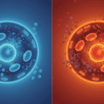
Your heart keeps beating through chemotherapy. The problem is, many cancer drugs are indiscriminate killers. They attack tumour cells brilliantly, but they also pump your cardiac myocytes full of reactive oxygen species, those unstable molecules that corrode cellular machinery from the inside. Some hearts survive this chemical onslaught better than others. New research suggests tiny cellular packages called extracellular vesicles might be acting as emergency shields, ferrying protective cargo directly to the cells that need it most.
What is oxidative stress in heart cells
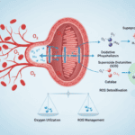
Oxidative stress happens when cells generate more reactive oxygen species than they can neutralise. Think of it like rust on metal, except it happens at the molecular level inside living cells. Your mitochondria produce these reactive molecules as a normal byproduct of energy production. Usually your cells manage this fine, using antioxidant enzymes like superoxide dismutase and catalase to convert dangerous radicals into harmless water and oxygen.
Heart cells are particularly vulnerable. Cardiac myocytes burn fuel constantly to generate the contractions that keep blood moving. They have high metabolic demands and packed-in mitochondria. When chemotherapy drugs enter the picture, they hijack this system. Drugs like anthracyclines intercalate into DNA and trigger electron leakage from the respiratory chain, flooding the cell with superoxide and hydrogen peroxide. The heart’s antioxidant defences get overwhelmed. The reactive molecules start damaging proteins, lipids, and DNA. Over time this triggers cell death or dysfunction.
What the research shows
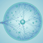
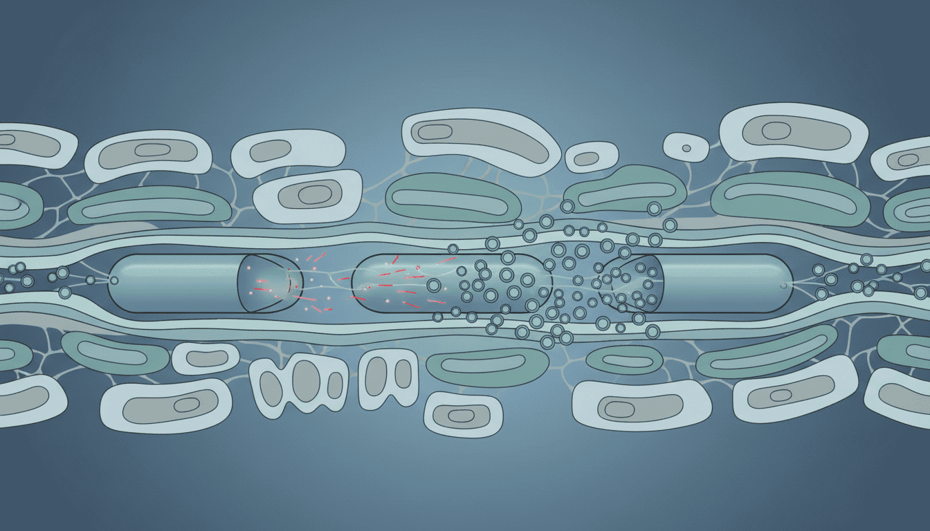
Scientists studying chemotherapy cardiotoxicity have observed something interesting happening at the cellular level. When heart cells face oxidative assault from cancer drugs, neighbouring cells and supporting tissues release extracellular vesicles in increased numbers. These vesicles are membrane-bound packages, ranging from 30 to 150 nanometres across, that bud off from cells and circulate through tissue fluid.
The protective cargo inside these vesicles is specific. Researchers have detected antioxidant enzymes packed into vesicles released by cardiac fibroblasts and endothelial cells in response to oxidative stress. More intriguingly, the vesicles contain signalling molecules and microRNAs that appear to upregulate the heart cell’s own defence systems. When cardiac myocytes take up these vesicles, they experience reduced oxidative damage and improved mitochondrial function compared to cells without this support.
The magnitude matters. Studies comparing hearts exposed to chemotherapy show that animals with robust extracellular vesicle signalling experience less contractile dysfunction and fewer signs of cardiomyocyte death. It is not a complete protection, but the difference is measurable and consistent across multiple experimental models.
Why cells need this defence
This protective system reflects evolution solving a fundamental problem: how do cells respond to neighbours in trouble? Your heart is an integrated tissue. A single cardiac myocyte cannot exist in isolation. It sits within a network of fibroblasts, endothelial cells, and immune cells. When oxidative stress hits, this network has mechanisms to communicate distress and coordinate defence.
Extracellular vesicles work as a chemical messenger system that works faster and more targeted than hormonal signalling. A fibroblast detecting oxidative stress in nearby myocytes can package protective molecules and release them within minutes. The receiving cell gets a direct boost to its antioxidant capacity without waiting for systemic signals to travel through the bloodstream.
This also makes biological sense from a redundancy perspective. If one cell’s defence systems fail completely, nearby cells amplify their own protection and share resources. It is cellular mutual aid built into the tissue architecture.
What affects extracellular vesicle protection
Age matters considerably. Older hearts produce fewer extracellular vesicles in response to oxidative stress, and the vesicles they do produce contain lower concentrations of protective cargo. This partly explains why chemotherapy cardiotoxicity increases with age. The tissue’s ability to mount this protective response deteriorates.
Existing cardiovascular disease impairs the system too. In hearts with prior hypertension or ischaemic damage, fibroblasts and endothelial cells show blunted vesicle release during oxidative stress. The tissue is already in a compromised state and cannot mobilise the defence as effectively.
Exercise training appears to enhance this protective capacity. Animal studies show that hearts conditioned through regular aerobic activity produce more functional extracellular vesicles at baseline and mount a stronger vesicle response to experimental oxidative stress. The mechanism is not entirely clear, but likely involves training-induced improvements in mitochondrial function and antioxidant enzyme expression in fibroblasts and endothelial cells.
Nutritional status influences the system as well. Hearts with adequate micronutrient intake, particularly minerals involved in antioxidant enzymes like copper and zinc, generate vesicles with higher protective capacity. Conversely, deficient states reduce the quality of the protective cargo being packaged.
What remains unknown
The specificity of vesicle targeting is still unclear. How does a cardiac myocyte preferentially take up vesicles designed to help it? What surface markers on vesicles allow them to dock with damaged cells rather than healthy neighbours? Researchers have identified some adhesion molecules involved, but the complete targeting mechanism remains a puzzle.
We also do not fully understand the limits of this protective system. At what concentration of oxidative stress does vesicle protection become insufficient? Does repeated or chronic chemotherapy exposure exhaust the tissue’s capacity to produce protective vesicles? Can you artificially enhance this response?
The relationship between inflammation and extracellular vesicle signalling deserves more attention too. Chemotherapy triggers both oxidative stress and immune activation in the heart. Do inflammatory signals amplify or suppress the protective vesicle response? Does timing matter, such as whether inflammation peaks before or after oxidative damage occurs?
Finally, we need better understanding of how extracellular vesicles interact with different chemotherapy drugs. Some agents might suppress vesicle production directly. Others might damage the protective machinery inside the vesicles. This drug-specific knowledge could identify which patients face highest risk.
The bigger picture here is that your cells are not isolated units passively accepting chemical damage. They are networked, communicating constantly, and able to mobilise collective defence when threats emerge. Extracellular vesicles represent one mechanism evolution has preserved for this tissue-level survival strategy. Understanding how to maintain or enhance this natural protection is an open question in cellular cardiology, and one that reaches beyond chemotherapy to any condition generating oxidative stress in the heart.

Matt Elliott is the editor of Redox News Today, an independent publication covering peer-reviewed research on cellular health, redox signalling, and related biomedical science.