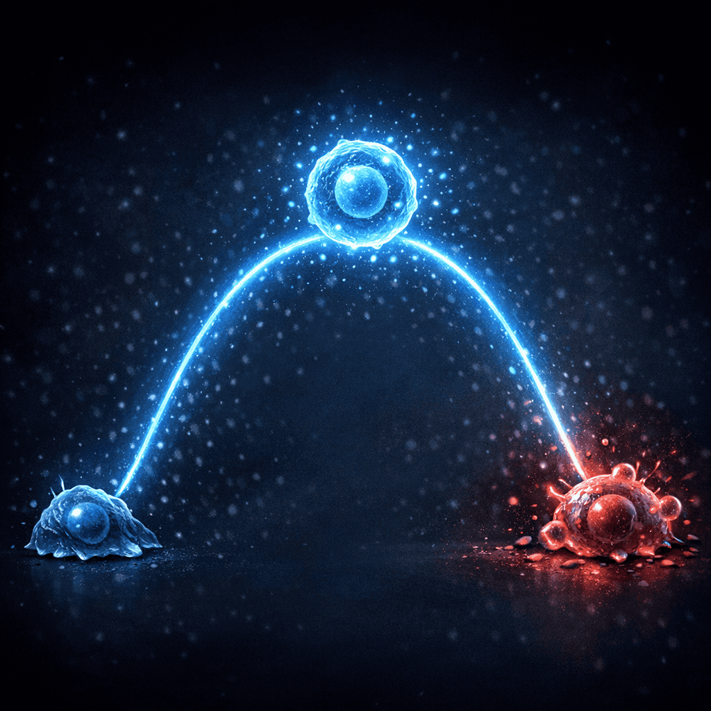
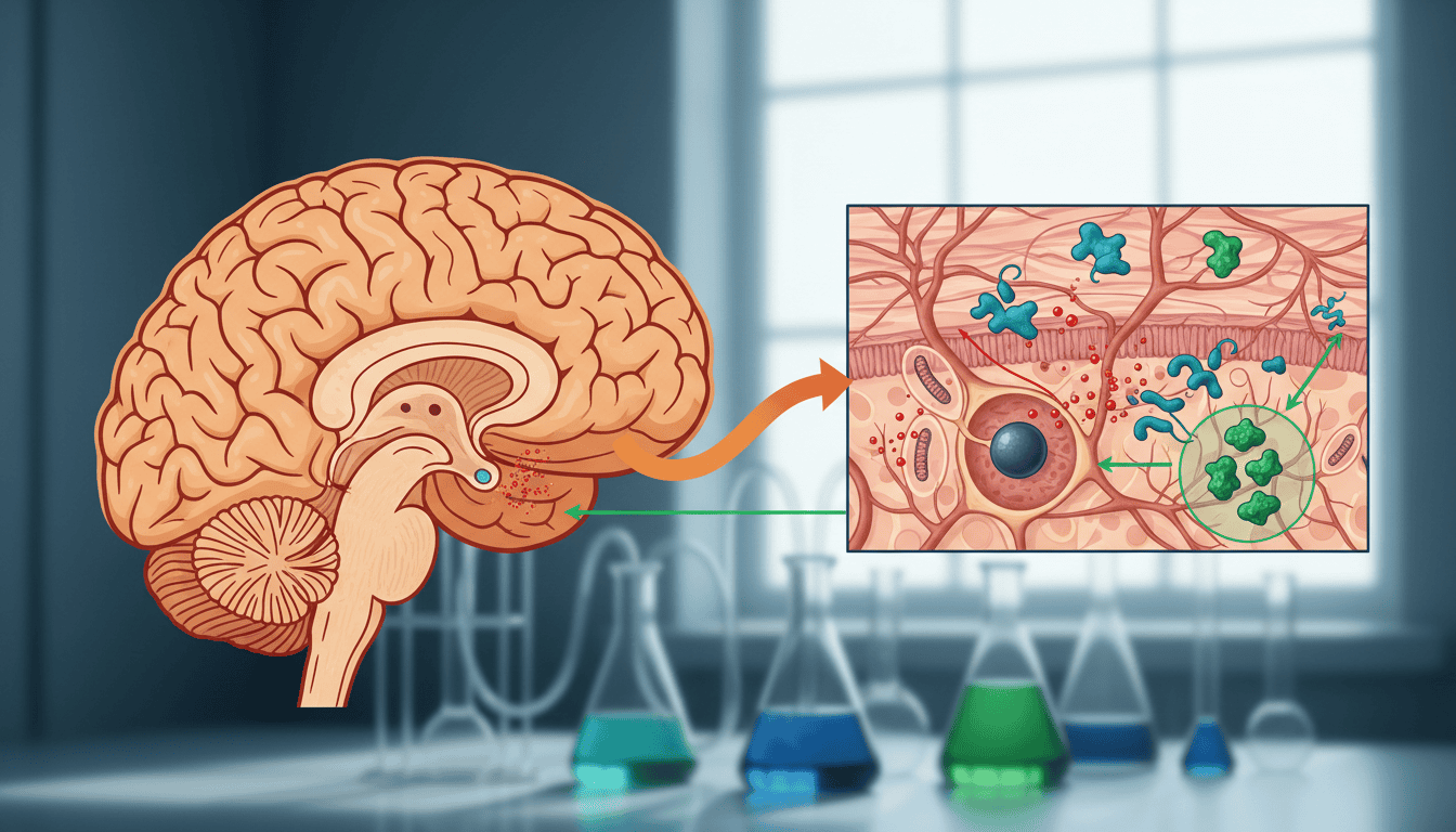
Your brain doesn’t like oxygen. Well, it needs oxygen to survive, but the byproducts of using oxygen are toxic. When a brain injury happens, this problem gets catastrophically worse. Cells are damaged, energy production falls apart, and reactive oxygen species flood the tissue like a chemical fire. Now researchers have found that a protein called TIGAR acts as a damage control system, redirecting cellular resources away from destruction and toward repair. Understanding how TIGAR works during injury could reshape how we think about brain recovery at the cellular level.
What is TIGAR
TIGAR stands for TP53-induced glycolysis and apoptosis regulator, which tells you almost nothing if you’re not a biochemist. Think of it differently: TIGAR is a metabolic traffic controller. When cells burn glucose for energy, they produce a molecule called pyruvate. Usually, pyruvate gets processed through multiple pathways. TIGAR sits at this junction and makes choices about where that pyruvate goes. In healthy cells, this is routine housekeeping. In injured brain tissue, it becomes survival strategy.
The protein emerged from cancer research initially, where scientists noticed it helped damaged cells survive. But its real significance became apparent when researchers examined what happens after traumatic brain injury. TIGAR doesn’t just redirect energy flow; it suppresses the production of dangerous free radicals and simultaneously supports antioxidant defence systems. It’s not a passive bystander in recovery. It actively shapes which protective mechanisms activate and which metabolic pathways dominate.
What the research shows
Studies comparing brain tissue after injury reveal that TIGAR expression increases dramatically in the hours and days following trauma. This isn’t random activation; the protein appears in precisely the regions where oxidative stress runs highest. When researchers experimentally reduced TIGAR levels in animal models of brain injury, something striking happened: the tissue showed worse oxidative damage, less effective energy metabolism, and slower recovery of neurological function.
The mechanism involves redirecting pyruvate toward the pentose phosphate pathway rather than complete oxidation. This pathway produces NADPH, a crucial molecule that feeds antioxidant systems like glutathione. By making this metabolic choice, TIGAR essentially tells the cell: “Stop trying to extract maximum energy right now. Prioritise defence.” The cells that make this shift survive better. Their oxidative stress decreases. Their recovery capacity improves. In some studies, blocking TIGAR entirely made injury outcomes measurably worse.
Brain tissue doesn’t exist in isolation, though. Astrocytes, the support cells surrounding neurons, also express TIGAR after injury, and they appear to take on additional burden to protect their neuronal neighbours. The protein coordinates a tissue wide response, not just individual cell survival.
Why cells need this
Brain injury creates a metabolic crisis. When cells lose blood supply or structural integrity, their mitochondria can’t function normally. This means oxygen gets used inefficiently, generating excess reactive oxygen species instead of useful energy. The cell simultaneously needs more energy to repair damage and is generating less of it while simultaneously poisoning itself with free radicals. This is an impossible situation that requires immediate metabolic reprogramming.
TIGAR solves this by essentially saying: “Abandon maximum energy extraction. Shift resources to antioxidant defence.” This sounds like giving up, but it’s actually optimal for survival. A cell that produces less ATP but maintains its defensive systems will outlast a cell desperately trying to extract maximum energy while drowning in oxidative stress. Evolution selected for this protein because injured brains that mount this response recover better.
The protein also appears to limit apoptosis, the programmed cell death pathway that gets inappropriately triggered after injury. By suppressing certain death signals while supporting survival mechanisms, TIGAR extends the window of recovery before cells pass the point of no return.
What affects TIGAR
Age matters. Older brains show slower TIGAR upregulation after injury, which might partially explain why recovery is typically slower in aged individuals. Young animals activate TIGAR robustly and rapidly; the response becomes sluggish with age.
Pre-existing metabolic stress also influences TIGAR function. Animals with poor metabolic health before injury show dampened TIGAR responses. This suggests that baseline cellular health determines how effectively the injury recovery system activates. Chronic stress, inadequate sleep, and poor metabolic function all appear to blunt this protective response in research models.
Temperature matters too. Cooling the brain after injury, a technique used clinically in some cases, appears to enhance TIGAR signalling and amplify its protective effects. Heat stress has the opposite effect. Even modest changes in brain temperature alter how effectively this protein operates.
Genetics play a role as well. Some individuals carry variants affecting TIGAR expression or function. These variants don’t necessarily cause disease in healthy individuals but may influence recovery trajectories after injury.
What remains unknown
Scientists still don’t fully understand the precise signals that activate TIGAR after injury. Something detects damage and rapidly increases TIGAR expression, but the sensing mechanisms aren’t completely mapped. Is it reactive oxygen species themselves? Changes in energy status? Specific damage signals from broken cell components? Probably all of these, but the hierarchy remains unclear.
The timeline of TIGAR activity also needs clarification. It rises quickly after injury, but how long should it stay elevated? When should it decrease? Is prolonged high TIGAR expression beneficial or does it eventually become counterproductive? Different types of brain injury might require different TIGAR profiles, but research hasn’t established optimal patterns.
Translation to humans presents another frontier. Most research happens in rodent models. How faithfully does this translate? Do human brains respond similarly? The recent development of improved neuroimaging techniques might finally allow researchers to observe TIGAR activity in living human brains, but we’re not there yet. Additionally, whether interventions that boost TIGAR would actually improve clinical outcomes remains completely untested.
The bigger picture
TIGAR represents something fundamental about cellular biology: damage forces metabolic choices. Cells don’t simply try to preserve themselves at any cost. They make strategic decisions about resource allocation based on threat assessment. This protein is part of that decision making apparatus. Understanding these mechanisms doesn’t cure brain injury, but it reveals how cells prioritise survival. It shows us that injury recovery isn’t just about repairing damage; it’s about metabolic recalibration. As research continues, TIGAR might become a target for optimising recovery, not through direct intervention but by understanding conditions that allow natural protective mechanisms to work more effectively.

Matt Elliott is the editor of Redox News Today, an independent publication covering peer-reviewed research on cellular health, redox signalling, and related biomedical science.