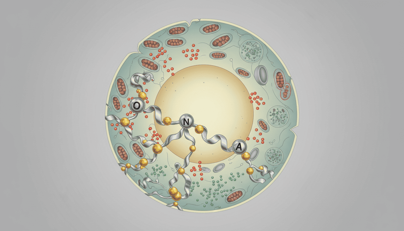
Your body contains roughly 30 billion fat cells, and they’re doing far more than storing energy. White adipose tissue is an active endocrine organ, constantly signalling to your brain, muscles, and immune system. Dump enough mercury into this system and something remarkable and troubling happens: the fat cells themselves become miniature sites of cellular damage, generating reactive oxygen species faster than your antioxidant defences can neutralise them. This oxidative stress then ripples through the fat tissue’s metabolism, potentially affecting how your body handles energy, inflammation, and glucose.
What is oxidative stress and how does mercury trigger it
Oxidative stress occurs when cells produce more reactive oxygen species (ROS) than they can handle. Think of ROS as molecular grenades: they’re generated naturally during normal metabolism, but in small quantities they actually serve useful purposes, helping cells signal and defend against pathogens. The problem emerges when ROS production spirals out of control. Mercury is particularly nasty at causing this imbalance. When mercury enters cells, it binds to proteins containing sulphur groups, particularly enzymes stuffed with cysteine residues. This binding disrupts crucial enzyme function while simultaneously triggering ROS generation as cells attempt to compensate. At the same time, mercury interferes with your antioxidant defence systems, specifically the glutathione (GSH) pathway, one of your cells’ primary ROS-neutralising mechanisms. The result is a two-pronged assault: more oxidative damage and fewer tools to fight it.
White fat cells are especially vulnerable to this attack. Unlike brown fat, which burns calories to generate heat, white fat stores energy and produces regulatory hormones. The mitochondria within white adipocytes generate ROS as a byproduct of normal energy production. Mercury exposure pushes this into overdrive, and the fat cells lack the robust antioxidant enzyme systems found in cells specialised for high metabolic activity.
What the research shows
Laboratory studies using cultured white adipocytes exposed to mercury reveal a consistent pattern: oxidative stress markers spike rapidly. Malondialdehyde (MDA), a product of lipid peroxidation, accumulates in the tissue. Protein carbonylation increases, indicating oxidative damage to cellular proteins. Glutathione levels plummet as cells deploy their antioxidant reserves. What’s particularly interesting is what happens to the fat cells’ metabolic machinery. Mercury exposure impairs adipogenesis (the process of fat cell development) and alters lipid metabolism. Some studies show increased triglyceride accumulation; others reveal impaired glucose uptake and mitochondrial dysfunction. The fat cells become metabolically confused, struggling to perform their normal functions while simultaneously fighting oxidative damage.
Gene expression studies show upregulation of inflammatory cytokines like TNF-alpha and IL-6, and markers of cellular stress response pathways activate. The MAPK and NF-kB signalling cascades, which orchestrate inflammatory responses, shift into high gear. Mercury also disrupts autophagy (the cell’s recycling process), preventing damaged components from being efficiently cleared away. In animal models exposed to environmental mercury levels, white adipose tissue shows increased oxidative stress markers compared to control animals, along with altered metabolic parameters and inflammation.
Why cells need functional fat tissue metabolism
White adipose tissue isn’t metabolically inert storage. It’s a metabolic powerhouse that produces hormones like leptin and adiponectin, which regulate hunger and whole-body glucose metabolism. Fat tissue also stores and mobilises fatty acids based on your body’s energy demands. When oxidative stress damages this tissue, the consequences extend far beyond the fat cells themselves. Inflammatory signals leak into the bloodstream, affecting distant tissues. Impaired fat mobilisation disrupts energy availability. Dysregulated hormone production confuses metabolic signalling throughout your body.
Evolution preserved this system because efficient fat storage and energy mobilisation are fundamental to survival. The oxidative stress defence mechanisms in your cells evolved to handle the normal ROS generated during metabolism. Mercury circumvents these evolutionary safeguards by generating ROS faster than cellular defences can manage, and simultaneously crippling those defences.
What affects mercury-induced oxidative stress in fat tissue
Individual susceptibility to mercury toxicity varies considerably. Genetic variations in glutathione S-transferase (GST) enzymes, which help process and eliminate mercury, create different baseline vulnerabilities. People with lower GST activity accumulate more mercury and experience greater oxidative stress. Nutritional status matters enormously. Selenium, zinc, and copper are cofactors for superoxide dismutase (SOD) and other antioxidant enzymes; deficiency in these minerals impairs your cells’ ability to handle ROS. Dietary antioxidant intake (vitamin E, vitamin C, polyphenols from food) influences how effectively cells manage oxidative stress, though the effect is modest compared to mercury’s damage potential.
Age influences the picture. Older cells generally produce more ROS and have less efficient antioxidant defence systems. Mercury exposure therefore tends to cause greater oxidative stress in ageing white adipose tissue. Chronic inflammation from other sources (poor diet, sedentary lifestyle, infection) creates a hostile backdrop that worsens mercury’s effects. Even alcohol consumption affects glutathione levels and antioxidant enzyme activity, potentially increasing vulnerability. Mercury speciation matters too: organic mercury compounds like methylmercury penetrate cells more readily than inorganic forms.
What remains unknown
Despite considerable research, fundamental questions persist. We don’t fully understand individual variation in mercury accumulation within fat tissue, or why some people’s adipocytes resist mercury-induced oxidative stress better than others. The long-term metabolic consequences of chronic, low-level mercury exposure remain somewhat opaque. Does oxidative stress in white fat tissue contribute to insulin resistance or metabolic syndrome in exposed populations? Some studies suggest yes, but causality remains difficult to establish in humans. We’re also unclear about the reversibility of these effects: if mercury exposure stops, how quickly do white adipocytes restore normal metabolic function and antioxidant capacity? Can cells recover fully, or does oxidative damage leave permanent marks? The interaction between mercury toxicity and other environmental stressors (other heavy metals, persistent organic pollutants) is understudied, despite the likelihood that real-world exposures involve multiple simultaneous threats.
Researchers are working on identifying biomarkers that reliably reflect mercury-induced oxidative stress in accessible tissues, which would allow better assessment of individual exposure and risk. Understanding which populations are most vulnerable based on genetics and nutritional status could inform targeted interventions.
This research reveals something fundamental about how cells respond to chemical threats. Your body has elegant defences against oxidative stress, but these systems can be overwhelmed. Mercury represents a particular threat because it disables defences while simultaneously increasing the damage they’re meant to prevent. White adipose tissue, far from being a passive energy depot, emerges as a frontline battleground where cellular toxicology plays out at the metabolic level.

Matt Elliott is the editor of Redox News Today, an independent publication covering peer-reviewed research on cellular health, redox signalling, and related biomedical science.