
Your mitochondria contain their own DNA, separate from the genetic material in your cell’s nucleus. But here’s the catch: mitochondria can’t manage this DNA alone. They rely on proteins made in the nucleus that must somehow travel across cellular space to reach them. Two proteins, MDM2 and TFAM, have emerged as key players in this intricate dance that determines whether your cellular power plants thrive or falter.
What is protein translocation to mitochondria
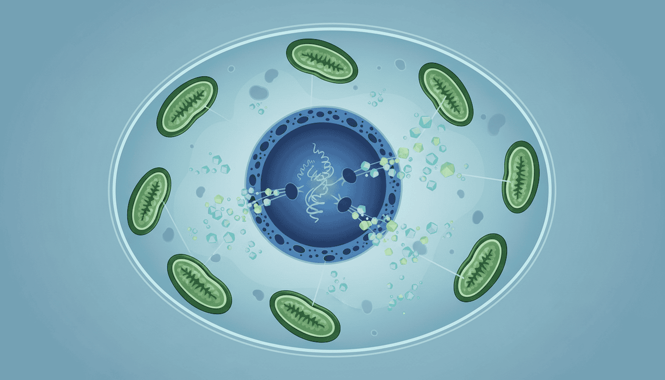
Mitochondria face a logistical challenge. These organelles need hundreds of different proteins to function, yet their own DNA only codes for 13 of them. The remaining proteins must be manufactured in the cell’s main protein factories, then transported across two separate membranes to reach their destination.
TFAM (Transcription Factor A, Mitochondrial) acts like a mitochondrial librarian. Once it arrives, TFAM binds to mitochondrial DNA and helps regulate which genes get switched on. Without enough TFAM, mitochondria struggle to produce the proteins they need for energy production.
MDM2 operates differently. Best known for its role in controlling cell division, this protein also shuttles between the nucleus and mitochondria. When MDM2 relocates to mitochondria, it influences how these organelles respond to cellular stress and energy demands.
What the research shows
Scientists have observed that the movement of these proteins isn’t random or constant. Cellular stress triggers a coordinated response where both MDM2 and TFAM increase their migration to mitochondria.
During periods of oxidative stress, researchers found that MDM2 accumulates in mitochondria within hours. This translocation coincides with changes in mitochondrial gene expression and metabolic output. The mitochondria essentially shift into a different operational mode.
TFAM translocation follows similar patterns but with different timing. Studies show that TFAM levels in mitochondria can fluctuate throughout the day, following circadian rhythms. When TFAM transport gets disrupted, mitochondrial DNA becomes unstable and energy production drops measurably.
Research teams have also documented that the efficiency of this protein transport system changes with age. Older cells show reduced ability to move both MDM2 and TFAM to mitochondria, correlating with decreased metabolic flexibility.
Why cells need this system
Evolution preserved this complex transport system for good reasons. Mitochondria originated as separate bacteria that merged with early cells billions of years ago. Over time, most mitochondrial genes transferred to the nucleus, creating this dependency relationship.
The system allows for sophisticated control. Nuclear proteins can sense the cell’s overall energy status, stress levels, and growth signals. By controlling which proteins travel to mitochondria and when, cells can rapidly adjust their energy production to match changing demands.
This flexibility proves essential during cellular stress. When cells detect damage or nutrient scarcity, they can quickly redirect mitochondrial metabolism by sending specific proteins to these organelles. It’s more efficient than waiting for mitochondria to slowly ramp up protein production using their limited genetic toolkit.
The dual control also provides a safety mechanism. If mitochondrial DNA gets damaged, nuclear proteins can compensate by increasing their transport and taking over some regulatory functions.
What affects protein translocation
Physical exercise consistently enhances the transport of both MDM2 and TFAM to mitochondria. Research shows that muscle contractions trigger signalling cascades that increase the efficiency of mitochondrial protein import machinery.
Nutritional status plays a major role. Fasting periods boost TFAM translocation as cells prepare mitochondria for increased fat burning. Conversely, high glucose conditions can suppress this transport, potentially contributing to metabolic dysfunction.
Temperature stress affects the system dramatically. Heat exposure and cold exposure both trigger increased MDM2 movement to mitochondria, though through different molecular pathways. This response helps mitochondria adapt their energy output to changing thermal demands.
Ageing systematically impairs this transport system. The protein machinery responsible for recognising and importing nuclear proteins becomes less efficient over time. This decline contributes to the metabolic changes observed in older organisms.
Certain medications and environmental toxins can interfere with protein translocation. Some antibiotics, for instance, disrupt mitochondrial import processes as an unintended side effect of their antimicrobial action.
What remains unknown
Scientists still can’t predict exactly which proteins will translocate to mitochondria under specific conditions. The signalling networks that control this process involve hundreds of interacting molecules, creating complexity that current research methods struggle to fully map.
The timing mechanisms remain mysterious. Researchers know that protein translocation follows circadian patterns, but they don’t understand how cells coordinate these rhythms with energy demands throughout the day.
Individual variation presents another puzzle. People show dramatically different efficiency in mitochondrial protein transport, but genetic studies haven’t identified all the factors responsible for these differences.
The relationship between protein translocation and disease development needs more investigation. While scientists observe disrupted transport in various conditions, they can’t yet determine whether this represents a cause or consequence of cellular dysfunction.
The story of MDM2 and TFAM illustrates how cellular biology operates through intricate partnerships rather than isolated components. These proteins reveal that mitochondria, despite their bacterial origins, have become deeply integrated into cellular regulatory networks. Understanding this integration helps explain why mitochondrial health depends not just on the organelles themselves, but on the cell’s ability to coordinate complex molecular traffic across internal boundaries.
Matt Elliott is the editor of Redox News Today, an independent publication covering peer-reviewed research on cellular health, redox signalling, and related biomedical science.


